Medical imaging depends on clear, dependable communication. Ultrasound Paper remains useful because it converts screen-based findings into physical clinical records. A printed image can travel with a patient file, support bedside discussions, and help explain results to families. In busy ultrasound rooms, staff can print selected frames within seconds. This simple step may improve continuity between sonographers, physicians, and patients.
High-quality Ultrasound Paper should produce sharp anatomical edges, balanced grayscale tones, and consistent contrast. These details matter when reviewing fetal structures, abdominal organs, or vascular findings. Thermal imaging paper also reduces dependence on liquid ink and routine cartridge replacement. That can simplify daily maintenance and reduce interruptions during examinations. Experienced imaging teams still check paper compatibility, printer settings, and storage conditions before regular use. A paper roll that fits poorly can create jams and waste valuable examination time. Small details matter.
Reliability has limits. Printed thermal images may fade under heat, sunlight, or prolonged pressure. They should not replace verified digital archives or approved medical records. Instead, they work best as practical reference copies and communication tools. This balanced approach supports professional judgment and responsible documentation. Users should select paper tested for their specific printer and clinical workflow. Manufacturer guidance, institutional policy, and local recordkeeping requirements deserve careful attention. No single product fits every department. That is the honest answer. When chosen thoughtfully, Ultrasound Paper can support clearer conversations, smoother workflow, and more accessible medical imaging records. Its value comes from dependable performance, not exaggerated promises.
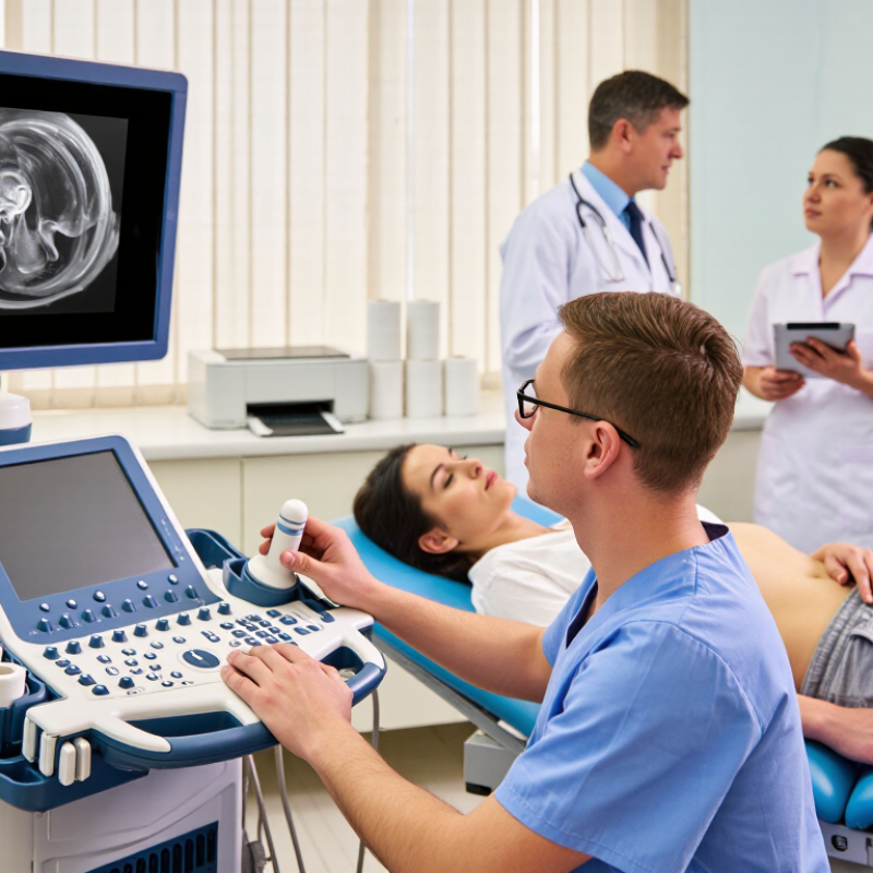
Ultrasound paper is a printed record produced by an ultrasound system. It usually uses heat-sensitive paper, not ink. The image appears through controlled thermal exposure. This process creates clear grayscale patterns from echoes, fluid spaces, and tissue boundaries. The U.S. Food and Drug Administration explains that diagnostic ultrasound uses sound waves rather than ionizing radiation. However, the paper records the displayed image, not the complete examination.
This distinction matters. A printed sheet may show a fetal profile, a cardiac valve, or a small cyst in a compact format. It is useful during consultations, bedside discussions, and patient handovers. The World Health Organization’s 2023 Global report on medical devices describes more than two million medical device types worldwide. That scale shows why simple, compatible consumables still matter. Yet ultrasound paper quality depends on thermal sensitivity, grayscale control, surface coating, and printer calibration. Small defects can hide fine borders.
Paper is not a perfect archive. Heat, sunlight, friction, and humidity can gradually weaken the image. Some facilities therefore keep digital studies as the primary record and use paper for quick reference. That approach follows practical imaging governance, but local retention rules still require review. A 2024 medical imaging workflow report noted that printed images remain common where rapid physical sharing is needed. The assumption that every paper printout is permanent deserves reconsideration. A clear image today may fade sooner than expected.
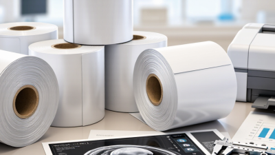
Ultrasound paper is heat-sensitive thermal paper used to print diagnostic images without ink or toner. Its common roll widths support different image formats, from compact reports to larger clinical printouts.
The chart shows common nominal roll widths used for medical thermal imaging paper: 110 mm, 112 mm, and 210 mm. Actual compatibility depends on the printer and imaging system specifications.
Ultrasound paper does not capture images by itself. A transducer sends sound waves into the body, while the imaging system converts returning echoes into grayscale data. The printer then transfers that image onto heat-sensitive paper, usually without ink. Each darker area represents stronger reflected signals, helping clinicians review tissue boundaries, fluid spaces, and measured structures.
Details matter. A clear print should show patient information, examination date, depth markers, and measurement lines. Fine contrast also helps preserve small differences between black, gray, and white areas. In daily imaging practice, technicians often check the printed image beside the monitor before releasing it. This simple comparison can reveal weak contrast or missing labels.
Paper is practical, but it is not flawless. It can fade under heat, sunlight, or high humidity. It may also curl after long storage. I think this limitation deserves more attention. A printed image should support clinical communication, not replace the original digital record. Staff should use suitable storage conditions and verify the patient identifiers carefully. One wrong label can create serious confusion. Good paper preserves visible evidence, but careful handling preserves its meaning.
Ultrasound paper remains useful in busy healthcare facilities because it turns an electronic scan into a visible, portable record. The Lancet Commission on Diagnostics reported in 2021 that nearly 47% of people worldwide have little or no access to diagnostics. In these settings, printed images can support referrals, bedside discussions, and follow-up when internet access is unstable.
It is practical. A clinician can mark a measurement, attach the image to a patient file, or hand it to a specialist within seconds. Paper also reduces dependence on screens during consultations. The World Health Organization’s 2023 guidance on medical equipment planning emphasizes reliable, maintainable tools, especially in resource-limited facilities. Ultrasound paper needs no battery. It is easy to store in folders and simple to replace.
Still, paper is not perfect. It can fade, curl, or carry an incorrect patient label. That risk demands careful verification before printing. The American College of Radiology recommends retaining diagnostic images in secure digital systems, so paper should support—not replace—DICOM archives and approved records. Staff should check contrast, thermal compatibility, and image size before purchasing supplies. Small details matter. A poor print can hide a fetal measurement or blur a vascular boundary. This is where routine quality checks remain essential.
Comparing ultrasound paper with digital imaging records requires more than a storage decision. In a busy ultrasound room, a printed image can move with the patient, even when network access fails. Clinicians can mark a measurement beside the image. Patients can also understand paper more quickly during bedside discussions. That matters. However, paper is easily folded, misplaced, or exposed to moisture. A clear label and controlled filing process remain essential.
Digital imaging records
Digital imaging records offer faster retrieval and stronger continuity across departments. Clinicians can compare today’s scan with older studies without searching through cabinets. Digital systems also support audit trails, backups, and structured reporting. The World Health Organization’s Global Strategy on Digital Health 2020–2025 identifies interoperability, privacy, and governance as essential requirements. These safeguards need daily maintenance, not just software installation. The U.S. Department of Health and Human Services recorded 725 large healthcare data breaches in 2023. Digital convenience therefore carries measurable security responsibilities.
A practical workflow
A practical workflow may use both formats. Paper can support immediate communication and temporary bedside reference. Digital records should remain the authoritative clinical archive. Each printed image needs patient identification, examination date, and retention rules. Still, this hybrid approach can create duplicate records. Staff may forget to scan annotations. Digital files can also become inaccessible during outages. Local audits, staff training, and patient feedback should guide the final choice. The process will not be perfect. That is precisely why it needs review.
Choosing ultrasound paper involves more than comparing prices. The paper must match the printer, imaging system, and clinical workflow. Check the equipment manual for the required size, coating, and thermal sensitivity. Small mismatches can produce faded images, uneven printing, or paper jams.
Image clarity matters during consultations and follow-up visits. A suitable surface should reproduce subtle shades and fine anatomical details without excessive glare. Ask for sample sheets before placing a large order. Compare skin tones, tissue boundaries, and small labels under normal examination-room lighting. The result may look acceptable on a screen but weak on paper.
Handling also affects reliability. Paper should feed smoothly, resist smudging, and remain readable after storage. Consider humidity, shelf life, packaging, and whether staff can separate sheets quickly with gloved hands. Clean storage matters. Dust can damage rollers and reduce print quality.
Patient records create another concern. Choose paper that supports clear identification, accurate dates, and dependable archiving. Review applicable healthcare documentation requirements in your region. Paper quality alone cannot guarantee long-term preservation. Folders, temperature, and light exposure also matter.
A practical test often reveals more than a product description. Record print speed, noise, curling, and waste during a busy clinic session. I have found that a slightly higher-cost option may reduce reprints. Still, assumptions can fail. Different printers may react differently, so local testing deserves attention before routine use.